Monthly Archives: March 2008

archivesectioncatbanner

Histology: Cardiac Muscle

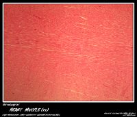
Cardiac muscle is the muscle of the heart. Cardiac muscle resembles skeletal muscle partially. It consists of short cylindrical muscle fibers, which branch and anastomose with each other. Each fiber contains a single oval centrally placed large nucleus. Some cells … Read More …


Posted in Year 1 Histology | Tagged | Leave a comment

Histology: Esophagus

The esophagus is a muscular tube that rapidly propels the food from pharynx into the stomach. It is about 25 cms long. Its wall includes all the layers included in the general plan of G.I.T. The mucous membrane is thrown … Read More …


Posted in Year 1 Histology | Tagged | Leave a comment

Histology: Blood Vessels

Large Blood Vessels Both the Aorta and Vena Cava have three layers. Tunicae intima, media, and adventitia. Aorta The Aorta is classified under elastic arteries (conducting arteries). They have (40-70) fenestrated membranes (Concentric layers of elastic membranes) at the tunica … Read More …


Posted in Year 1 Histology | Tagged | Leave a comment

Histology: Cartilage

Cartilage is a specialized dense connective tissue. Functions: (i) It bears the weight of the body and plays an important role during the growth in length of long bones. (ii) Gives support to developing organs. (iii) Cartilage helps in withstanding … Read More …


Posted in Year 1 Histology | Tagged | Leave a comment

Histology: Spleen

Spleen contains large amount of lymphatic tissue which filters blood instead of lymph. The spleen is surrounded by a connective tissue capsule which in lower animals contains smooth muscle fibers and elastic fibers. The outer surface of capsule is covered … Read More …


Posted in Year 1 Histology | Tagged | Leave a comment

Histology of respiratory system: Lung

LUNGS Histology of Bronchi: Primary, secondary, tertiary Epithelium – respiratory epithelium – columnar cells have gradual decrease in height, cilia & goblet cells. Lamina propria – gradual decrease in thickness & increase in number of elastic fibres. Glands – mucous … Read More …


Posted in Year 2 Histology | Tagged | Comments Off on Histology of respiratory system: Lung

Histology of Respiratory system: Trachea

TRACHEA Histology of Trachea Epithelum – respiratory epithelium (pseudo stratified columnar epithelium with goblet cells). Lamina propria – gradual decrease in thickness & increase in number of elastic fibres. Glands – mucous & serous glands. Skeletal connective tissue – C … Read More …


Posted in Year 2 Histology | Tagged | Comments Off on Histology of Respiratory system: Trachea

Histology: Skeletal Muscle

Structure of skeletal muscle. With haemotoxylin and eosin stain, skeletal muscle fibers are seen as highly easinophilic cylinders with multiple peripheral nuclei taking up a basic stain. Each fiber is surrounded by an outer limiting membrane called sarcolemma. The cytoplasm … Read More …


Posted in Year 1 Histology | Tagged | Leave a comment