Structure of skeletal muscle.
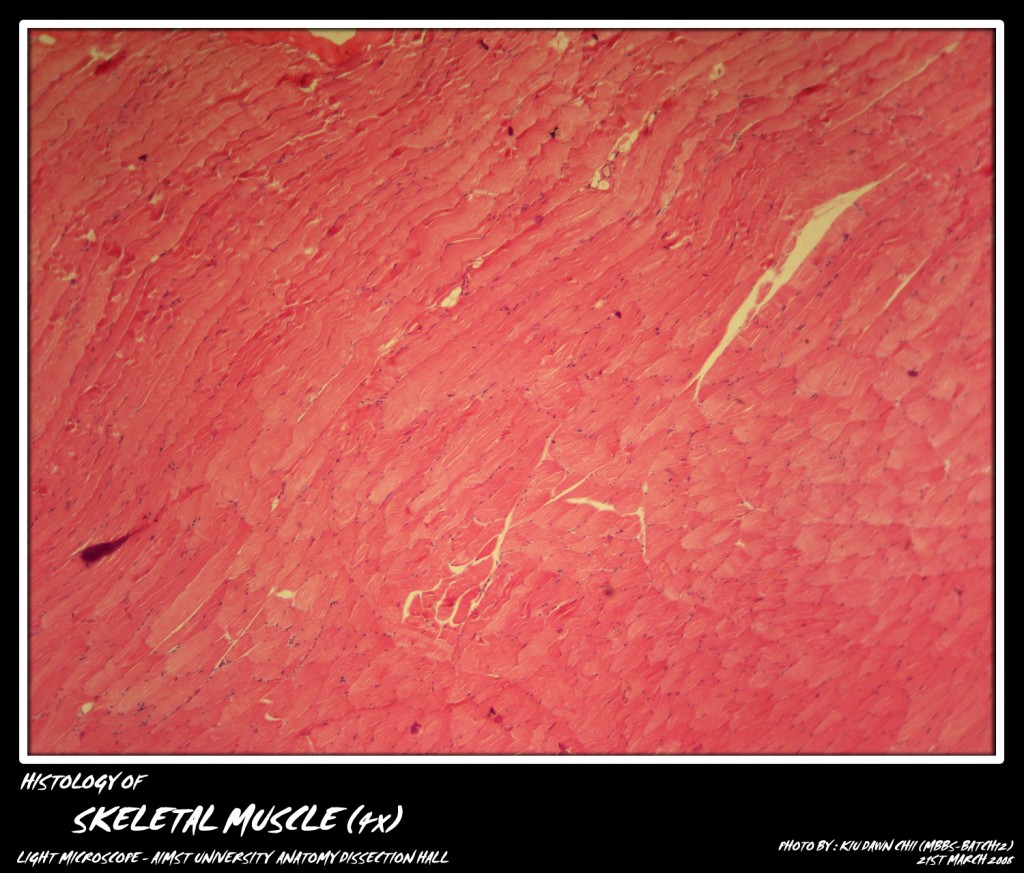
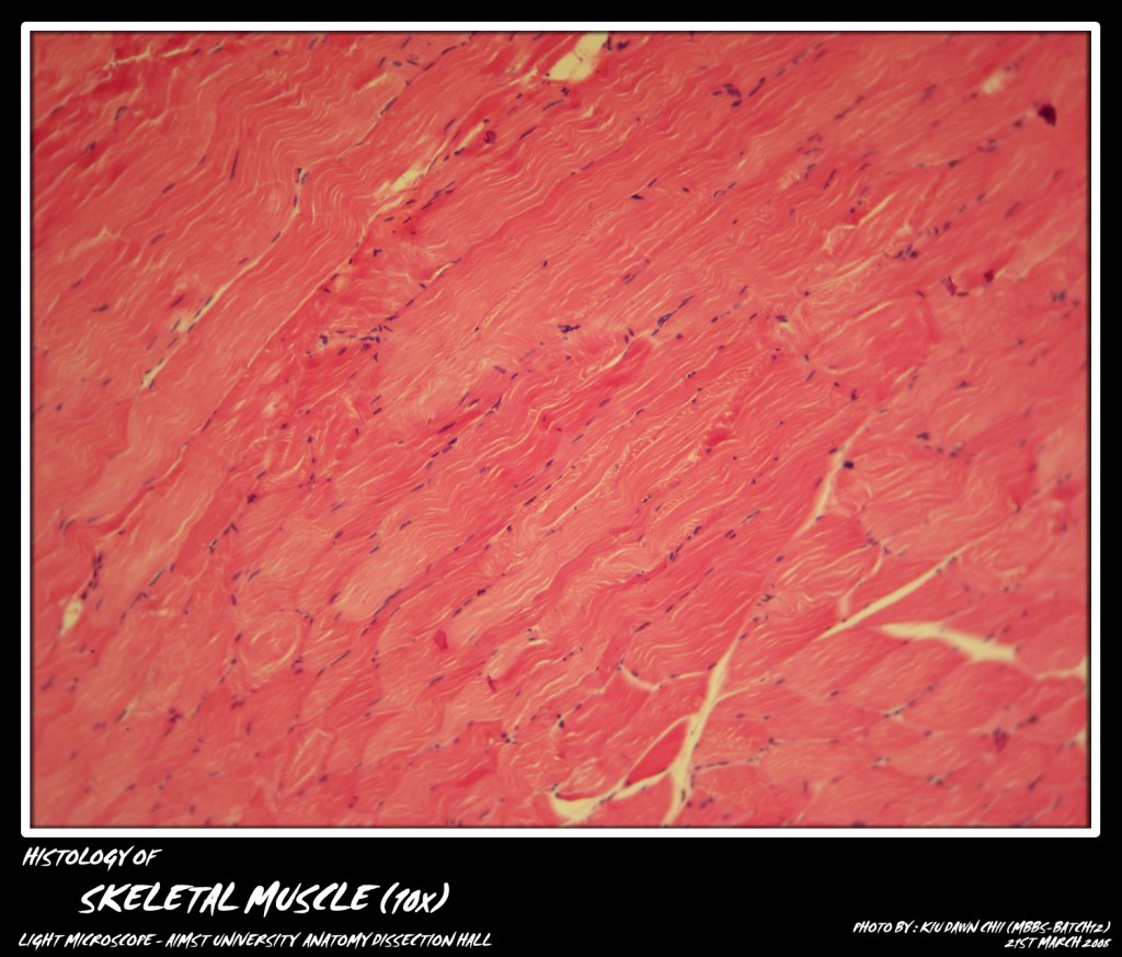
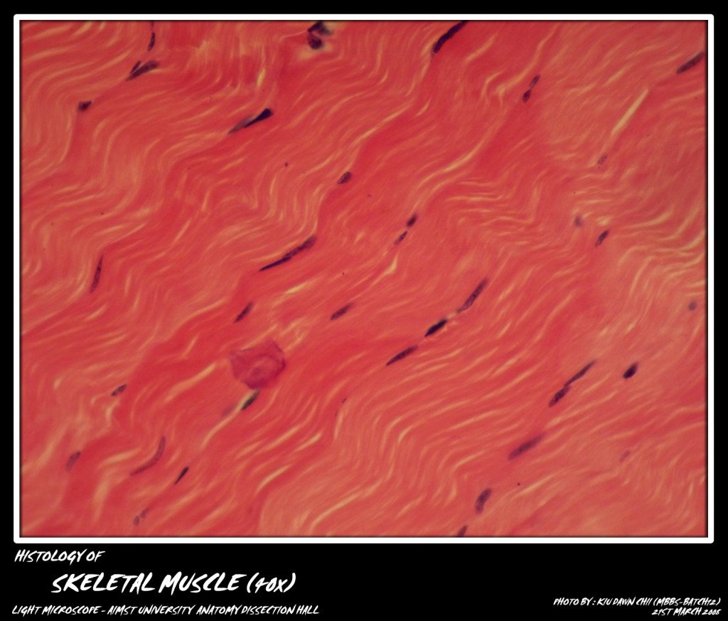
With haemotoxylin and eosin stain, skeletal muscle fibers are seen as highly easinophilic cylinders with multiple peripheral nuclei taking up a basic stain.
Each fiber is surrounded by an outer limiting membrane called sarcolemma.
The cytoplasm or sarcoplasm contains extensive sarcoplasmic reticulum, mitochondria and glycogen granules as well as large number of longitudinally running myofibrils. Under high power, the myofibrils depict alternating dark and light bands. These dark and light bands in all the myofibrils are “in register”, providing a continuous cross striations in the muscle fiber. These well marked transverse/cross striations are a diagnostic and characteristic feature of the striated muscle.
The micro-photograph shows the Skeletal Muscle at various magnification.
Each of the dark and light bands are intersected by lines:
Dark band or A band contains a light zone called the H zone. Light band or I band is similarly bisected by a dark transverse line the Z line. The functional unit of a muscle fiber is known as the sarcomere which is the segment between two successive Z lines, and therefore includes one A band and half of two contiguous I bands. During phases of contraction length of ‘A’ band remains constant while length of I band is greatest in stretched muscle, shorter in resting muscle and shortest in contracting muscle. The nuclei of skeletal muscle fibers are oval, multiple and peripheral. These are mostly seen beneath the sarcolemma at the periphery of the fiber. Some nuclei appearing in the central part of the fiber belong to the connective tissue around the muscle fibers.
Adapted from : http://myaimst.net/mbbsb12/photo/histo/yr1histo/muscle.html
Micro-photograph taken at AIMST University Anatomy Dissection Hall during Histology class, using Canon A40 camera over light microscope.


