Peripheral Nerve Fibers
A peripheral nerve fiber is an axon/Dendron with its covering i.e. myelin sheath and neurilemma. These fibers are myelinated. Each fiber consists of:
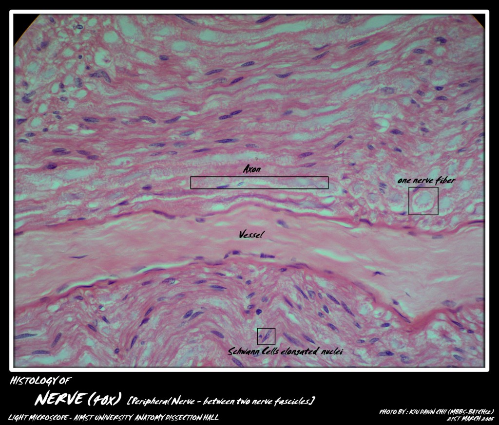
(i) A central axon/axis cylinder with azoplasm and neurofibrils contained within the axolemma.
(ii) Myelin sheath is composed of phospholipids, interrupted at intervals along the length of the fiber. It is stained by osmic acid and not by H & E stain.
(iii) Thin neurilemma sheath is present outside the myelin sheath. The cells of neurilemma are also known as Schwann cells, which are neuroectodermal in origin. At the points of interruption of myelin sheath the neurilemma comes into intimate contact with the axon and such areas are known as Nodes of Ranvier. The impulse passes from one node to the next node.
(iv) Endoneurium is a thin connective tissue layer of mesodennal origin. It supports the nerve fibers. The potential space between neurilemma and endoneurium contains tissue fluid for the nourishment of the nerve fiber.

Micro-photograph of Peripheral Nerve (showing between two nerve fascicles) under light microscope magnification 40x

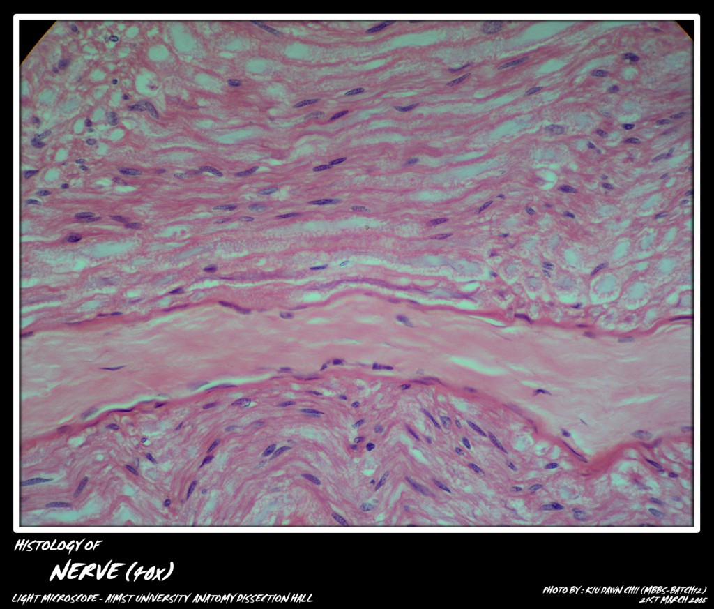
Micro-photograph of Peripheral Nerve (showing between two nerve fascicles) under light microscope magnification 40x
Adapted from: http://myaimst.net/mbbsb12/photo/histo/yr1histo/nervetissue.html
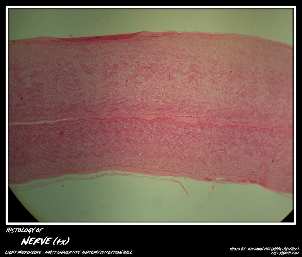
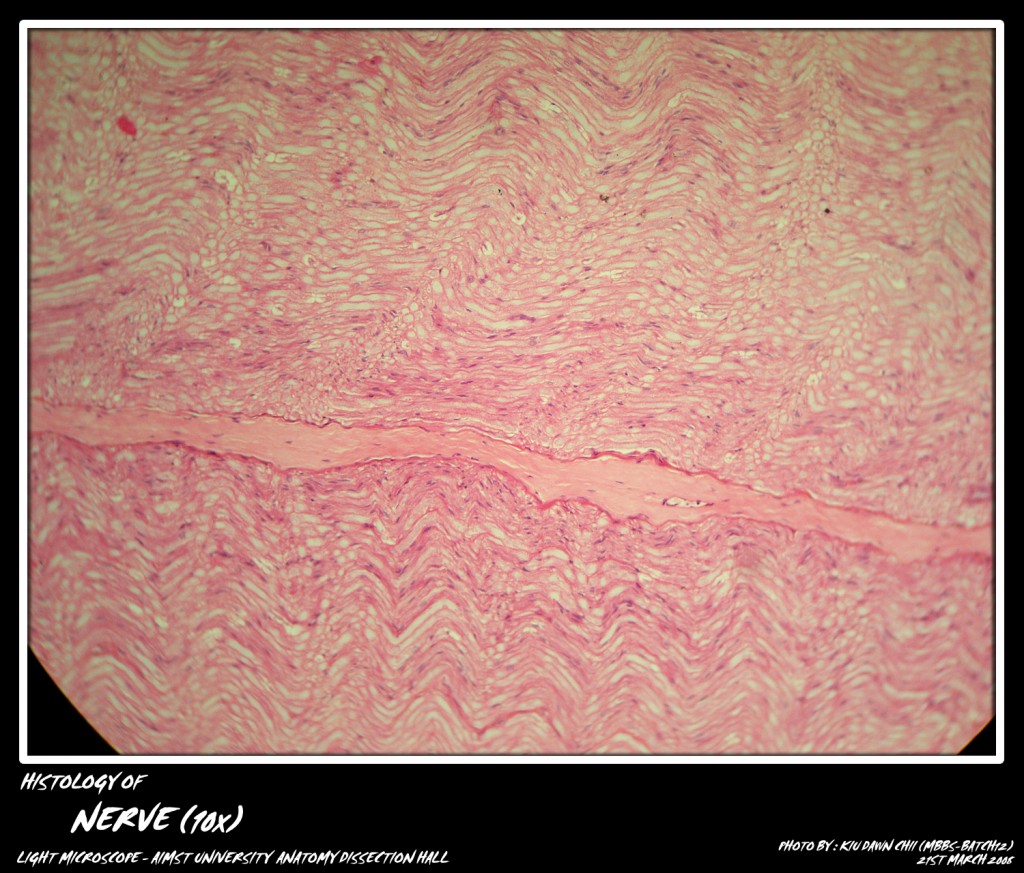
Micro-photograph taken at AIMST University Anatomy Dissection Hall during Histology class, using Canon A40 camera over light microscope.


