
BLOOD VESSEL
Classification of blood vessels based on the structure
- Arteries
- Elastic, Large or conducting arteriesConduct blood away from the heart.
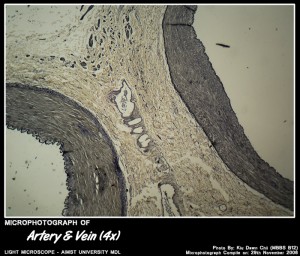
Micro-photograph of Artery VS Vein under Light Microscope magnification 4x
Important in maintaining constant pressure in arterial system
- Muscular arteries or medium sized arteriesThey distribute the blood to the tissues
- Elastic, Large or conducting arteriesConduct blood away from the heart.
- Veins
- Large veins
- Medium sized veins
Wall of the blood vessels
Typically having three concentric layers.
- Tunica intima – innermost layer
- Tunica media – middle layer
- Tunica adventitia (externa) – outermost layer
Histology of elastic arteries
- Tunica intima – thicker than in muscular arteries
. Endothelium rests on thin basal lamina
. Internal elastic lamina may be present between intima & media, but hard to distinguish because of abundant elastin fibres in media. - Tunica media – contains abundant elastin as concentrically arranged interspersed with smooth muscle fibres.
- Tunica adventitia – thin relative to vessel diameter, contains elastic & type I collagen fibres
,Vasa vasora are seen. - e.g. – aorta
Histology of Muscular or distributing arteries
- Tunica intima – Contains typical endothelium & subendothelial connective tissue.
Prominent internal elastic lamina appears as wavy, refractile line between intima & media. - Tunica media – Thick , Up to 40 layers of smooth muscle fibers.
Collagen, elastic fibres & proteoglycan vary. - Tunica adventitia – Relatively thin & contains mostly collagen fibres
Histology of Large veins
Tunica intima – Well developed & include thick layer of subendothelial connective tissue.
Extensions of intima protrude into lumens of large veins as valves.- Tunica media – several layers of smooth muscle cells & abundant reticular & collagen fibres. Elastin is sparse.
- Tunica adventitia – best developed in large veins contains abundant collagen & longitudinal bundles of smooth muscle that strengthen vessel to prevent distension.
- Eg.- Vena cava
Histology of Small & medium sized veins
Narrower than large veins & have thin walls.
- Tunica intima -Typical endothelium, less subendothelial tissue. fewer valves than large veins, no internal elastic lamina.
- Tunica media -Thin relative to vessel diameter. Few elastic fibres.
- Tunica adventitia – Relatively thick, but unlike large veins contains little of any muscle.
Mostly collagen. - e.g.– saphenous, hepatic, portal etc.
Micro-Photograph of Blood Vessels
Below are blood vessels at various magnification.
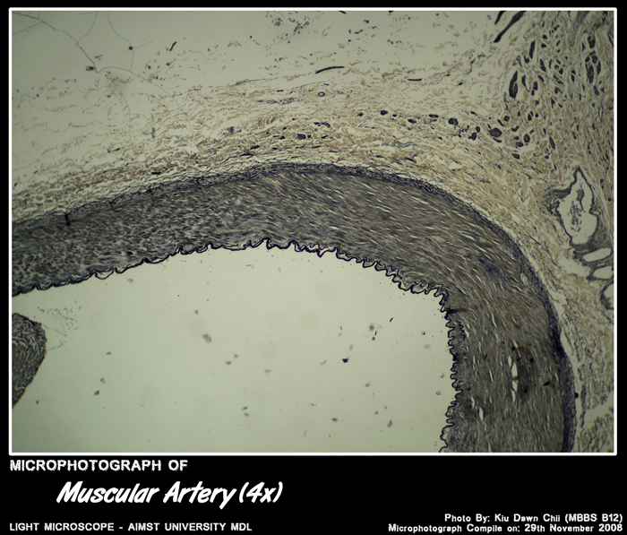
Micro-photograph of Muscular Artery under Light Microscope magnification 4x
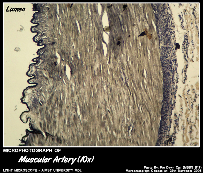
Micro-photograph of Muscular Artery under Light Microscope magnification 10x

Micro-photograph of Aorta – Large Elastic Artery under Light Microscope magnification 10x
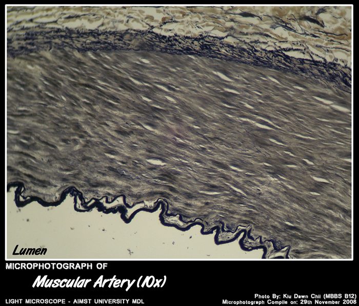
Micro-photograph of Muscular Artery under Light microscope magnification 10X

Micro-photograph of Artery VS Vein under Light Microscope magnification 4x
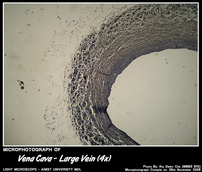
Micro-photograph of Vena Cava – Large Vein under Light Microscope magnification 4x
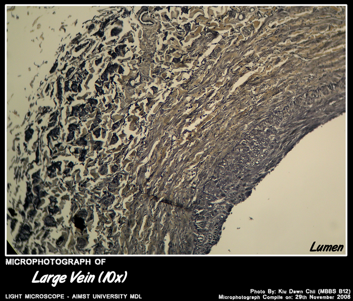
Micro-photograph of Large Vein under Light Microscope magnification 10x
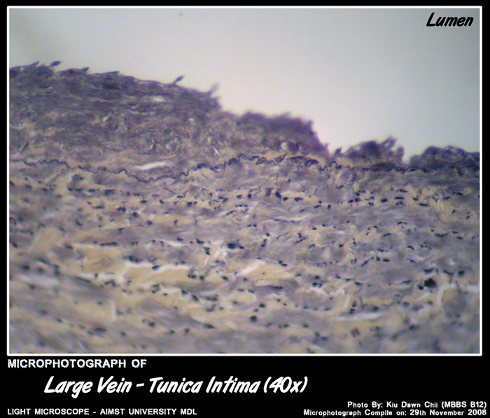
Micro-photograph of Large Vein – Tunica Intima under Light Microscope magnification 40x

Micro-photograph of Large Vein – Tunica Adventitia under Light Microscope magnification 40x
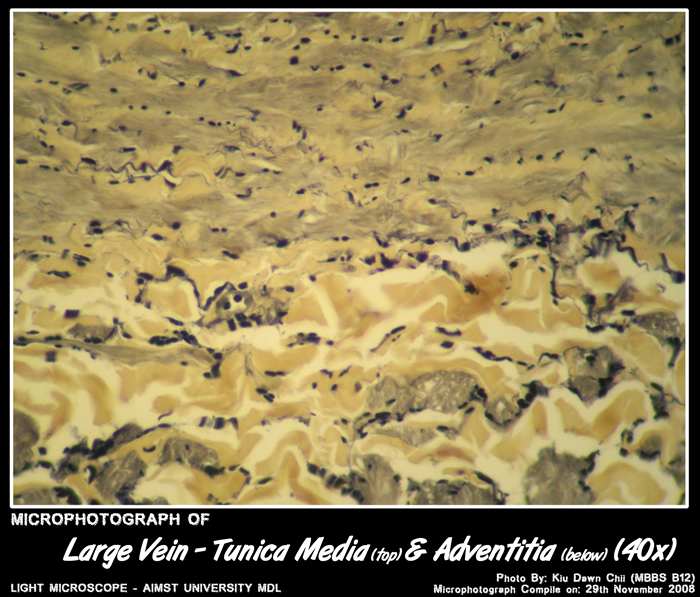
Micro-photograph of Large Vein – Tunica Media (top) and Adventitia (below) under Light Microscope magnification 40x
Adapted from: http://myaimst.net/mbbsb12/photo/histo/yr2histo/bloodvessels.html
Micro-photograph taken at AIMST University Multi Disciplinary Laboratory during Histology class, using Canon A40 camera over light microscope.