Striated Muscle
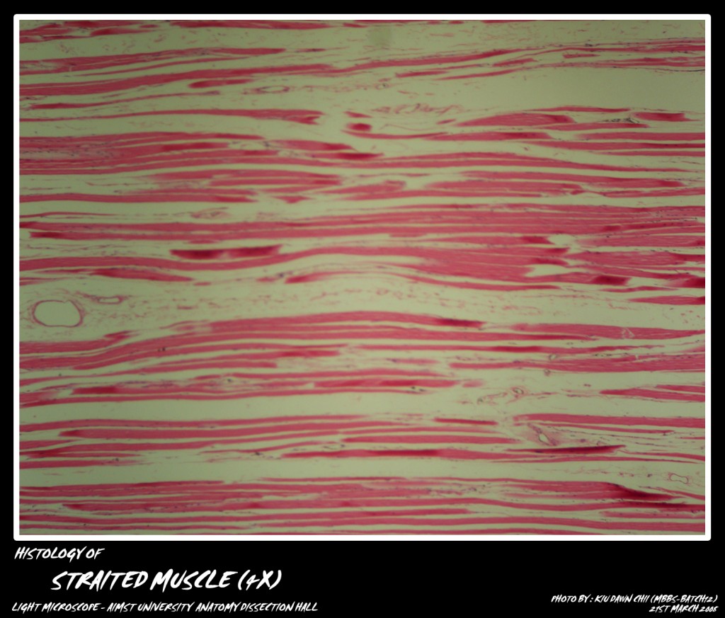
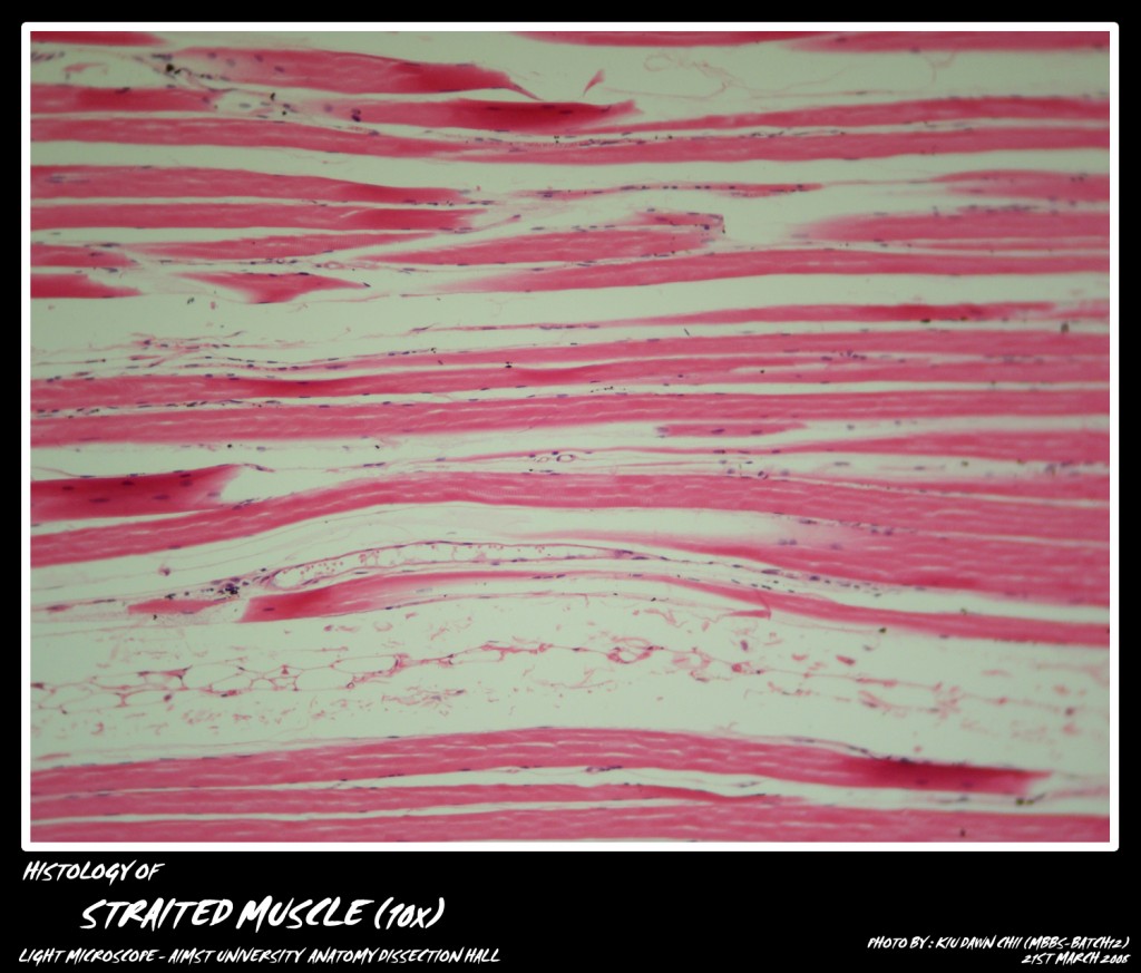
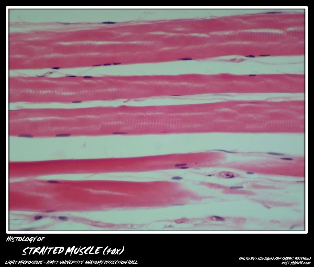
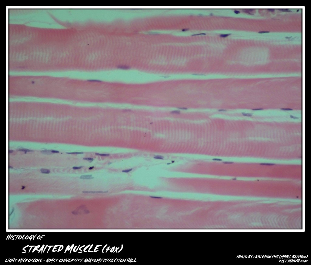
It is present in muscles of the limbs and trunk. The muscle as a whole is enclosed in a connective tissue layer called epimysium. Septa extend inwards from the epimysium dividing the muscle into various fasciculi. Thus each fasciculus is surrounded by perimysium from which extend fine sepia called endomysium that invest individual muscle fibers.
The micro-photograph shows the Striated Muscle at various magnification.
Adapted from: http://myaimst.net/mbbsb12/photo/histo/yr1histo/muscle.html
Micro-photograph taken at AIMST University Anatomy Dissection Hall during Histology class, using Canon A40 camera over light microscope.

