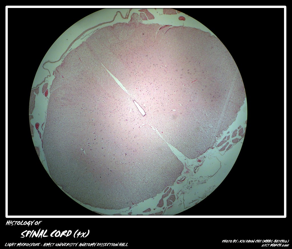
The spinal cord is comprised of a central canal surrounded by grey matter. Around this grey matter is the white matter.
Grey Matter
– Contains bodies of nerve cells.
– Has dendrites and parts of the axon.
– Contains protoplasmic astrocytes, oligodendroglia and microglia.
– Has numerous capillaries
White Matter
– Bodies of nerve cells are absent.
– Has most of the lengths of axon and dendrites.
– Contains fibrous astrocytes, oligodendroglia and microglia.
– Has fewer capillaries.
1. The central canal is seen as an oval cavity lined by columnar ciliated epithelium.
2. The large cells in the anterior horn depict multiple angles/corners, the angles representing the origin of its processes.
The grey matter reveals the neuroglial cells and lots of capillaries.
3. The peripheral white matter contains the fibers, neuroglia and fewer capillaries. In transverse sections the nerve fibers appear as hollow circles (myelin unstained) with central dots representing the axon.
Adapted from: http://myaimst.net/mbbsb12/photo/histo/yr1histo/nervetissue.html
Micro-photograph taken at AIMST University Anatomy Dissection Hall during Histology class, using Canon A40 camera over light microscope.


