
TRACHEA
Histology of Trachea
Epithelum – respiratory epithelium (pseudo stratified columnar epithelium with goblet cells).- Lamina propria – gradual decrease in thickness & increase in number of elastic fibres.
- Glands – mucous & serous glands.
- Skeletal connective tissue – C – shaped hyaline cartilage rings.
- Muscle – Smooth muscle named trachealis muscle.
The micro-photograph shows the Trachea at various magnification.
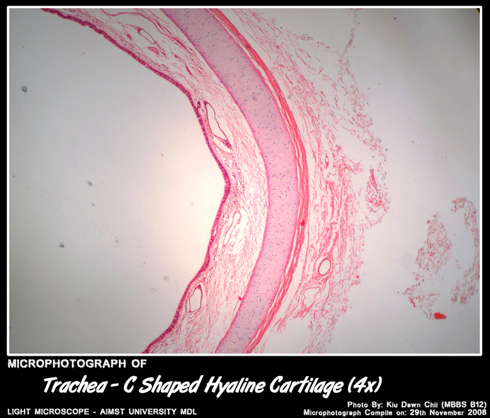
Micro-photograph of Trachea – C Shaped Hyaline Cartilage under light microscope magnification 4x
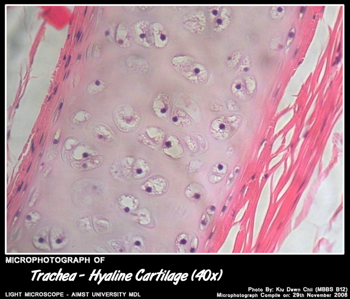
Micro-photograph of Trachea – Hyaline Cartilage under light microscope magnification 40x

Micro-photograph of Trachea – Trachealis Muscle under light microscope magnification 4x

Micro-photograph of Trachea – Trachealis Muscle under light microscope magnification 10x
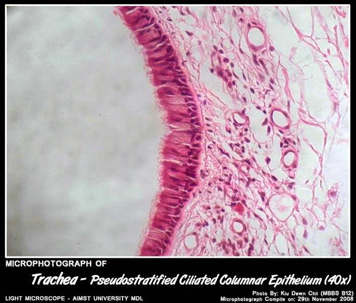
Micro-photograph of Trachea – Pseudostratified Ciliated Columnar Epithelium under light microscope magnification 40x
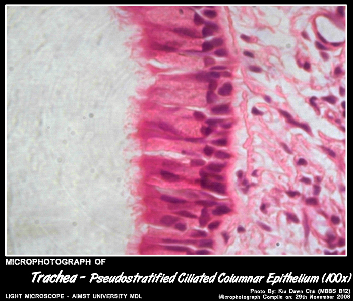
Micro-photograph of Trachea – Pseudostratified Ciliated Columnar Epithelium under light microscope magnification 100x
Adapted from: http://myaimst.net/mbbsb12/photo/histo/yr2histo/trachea.html
Micro-photograph taken at AIMST University Multi Disciplinary Laboratory during Histology class, using Canon A40 camera over light microscope.