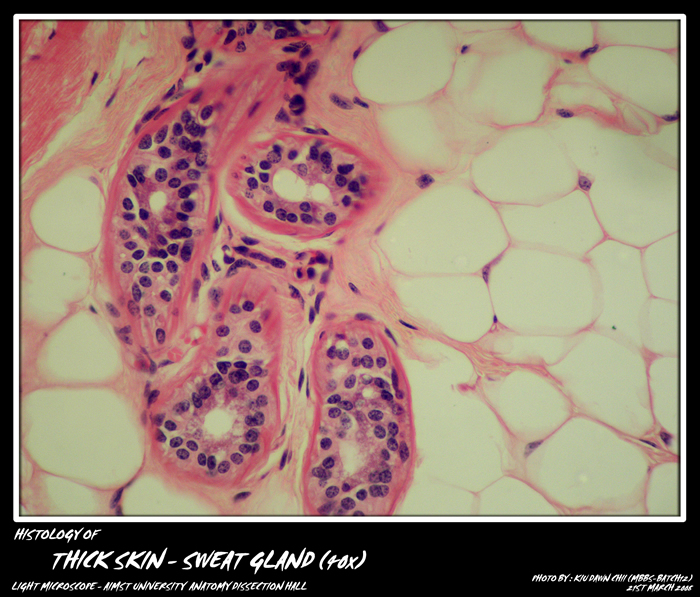
Skin is the largest organ of the body.It is a major barrier between the body and the external environment.
Main Functions of skin:
- Protection: It forms a protection layer against mechanical/thermal/chemical stress, infection
- Sensation: skin contains touch, pressure, pain and temperature receptors
- Thermoregulation: sweat glands, hair, and adipose tissue helps in thermoregulation
layers in skin:
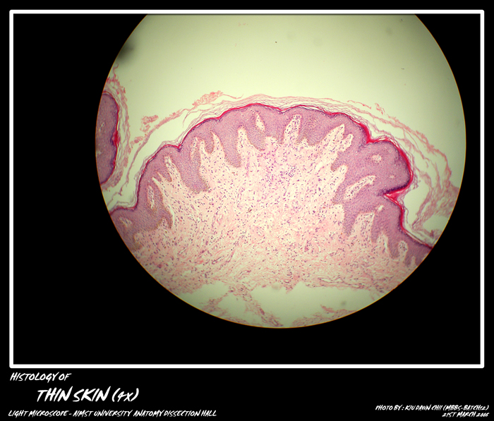
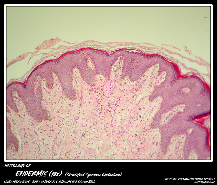
- Epidermis: formed by keratinised stratified squamous epithelium, it is the first barrier. The stratum lucidum layer is absent in thin skin.
- Dermis: thick connective tissue layer of the skin involves in sensation, protection and thermoregulation. It contains collagen and elastin fibres, and fibroblasts, macrophages and adipocytes, as well as nerves, glands and hair follicles.
The superficial region or called papillary dermis (have small projections called dermal papillae) which contains loose connective tissue with many capillaries, Meissners corpuscles (touch receptors), as well as free nerve endings.
The deeper region or called reticular dermis, is a layer of dense irregular connective tissue, which contains collagen and elastin, which give skin its strength and extensibility.
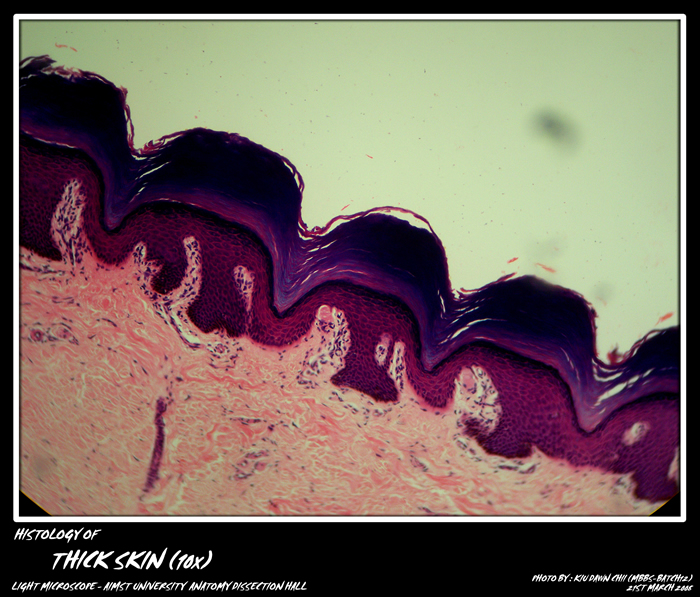
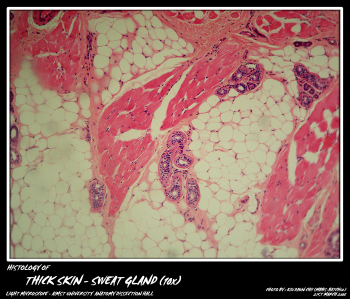
Thick skin found in areas where there is a lot of abrasion such as fingertips, palms and the soles of feet, does not contain hairs, sebaceous glands, or apocrine sweat glands. The dermis layer of thin skin is thicker than of thick skin. - Hypodermis: is a layer after dermis which contains mainly adipose tissue and sweat glands.
layers in skin
Cells in the dermis and epidermis.
- Keratinocytes or prickle cells makes up the majority of the cells (about 90%) in the epidermis.
- Melanocytes are found in the basal layer, are melanin producing cells.
- Langherhans cells are found in the stratum spinosum layer of the epidermis. They are irregularly shaped dendritic cells, facilitating skin allergic reactions.
- Merkel Cells are granular basal epidermal cells, attached to non-myelinated nerve ending, which are sensitive to touch.
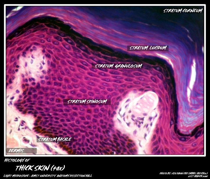
Micro-photograph of Epidermis layers of thick skin under light microscope magnification 40x (Showing stratum basale, stratum spinosum, stratum granulosum, stratum lucidum and stratum corneum)
Skin by nature of its barrier function, are constantly subject to wear and tear, hence constant proliferation of cells in the bottom layer (stratum basale), changing their appearance as they move up to the top layer to replace the lost cells.
Cell layers in the epidermis.
- Stratum Basale or stratum germinosum is a single layer of cells at the bottom, closest to the dermis, is a layer of active cell proliferation.
- Stratum Spinosum is the prickle cell layer of around 8-10 layers of cells, contains desmosomes (holds the intergrity between cells) and keratin (intermediate fillaments).
- Stratum Granulosum is a granule cell layer of around 3-5 layers of cells, they start to lose their nuclei and cytoplasmic organelles as they move up.
- Stratum Lucidum a thin transparent layer between the stratum granulosum and stratum corneum.
- Stratum Corneum is keratinised squamous layer of dead cells. They are flattened scales, or squames, filled with densely packed keratin.
Adapted from : http://myaimst.net/mbbsb12/photo/histo/yr1histo/epithelium.html
Micro-photograph taken at AIMST University Anatomy Dissection Hall during Histology class, using Canon A40 camera over light microscope.

