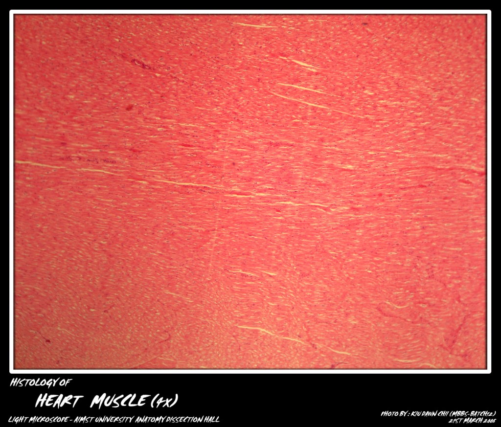
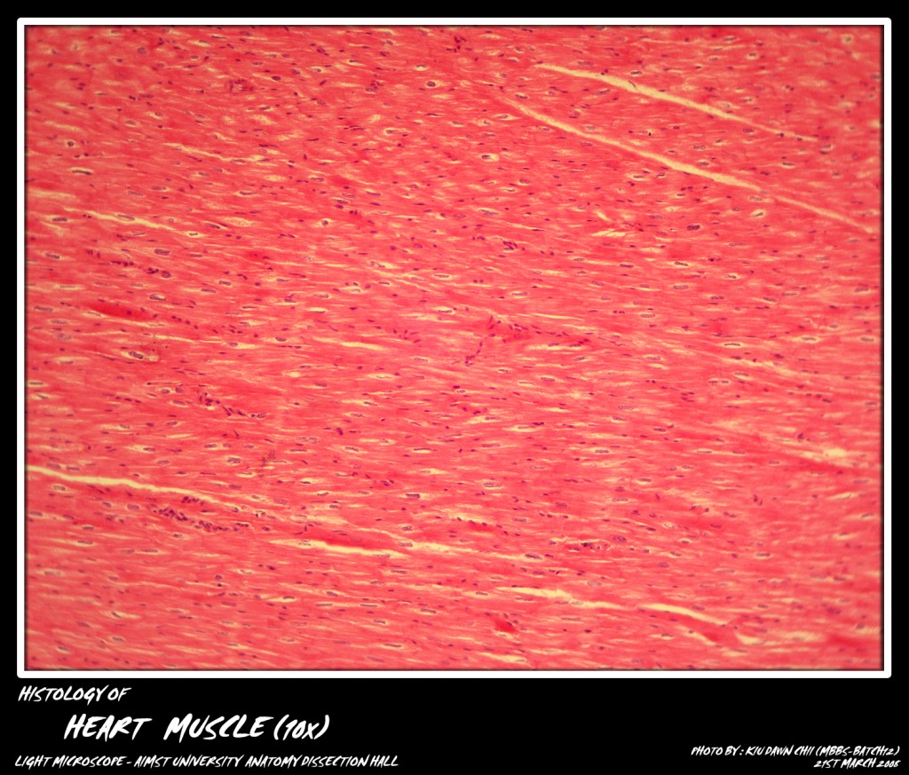
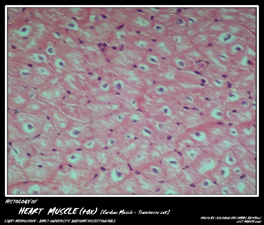
Cardiac muscle is the muscle of the heart. Cardiac muscle resembles skeletal muscle partially. It consists of short cylindrical muscle fibers, which branch and anastomose with each other. Each fiber contains a single oval centrally placed large nucleus. Some cells show a perinuclear clear space. The transverse striations are present but are not as conspicuous as in skeletal muscle.
The muscle fibers are joined together by surface specialisations known as intercalated dics. These intercalated/intercalary discs or junctional complexes appear as zigzag transverse lines and are caused by the apposed plasma membranes of the two fibers. These intercalated discs are better visible by silver stains. The spaces between the branchings of muscle fibers are occupied by fine connective tissue and blood cpaillaries. With increasing age, lipofuchsin pigment is deposited around the nuclei of the muscle fibers.
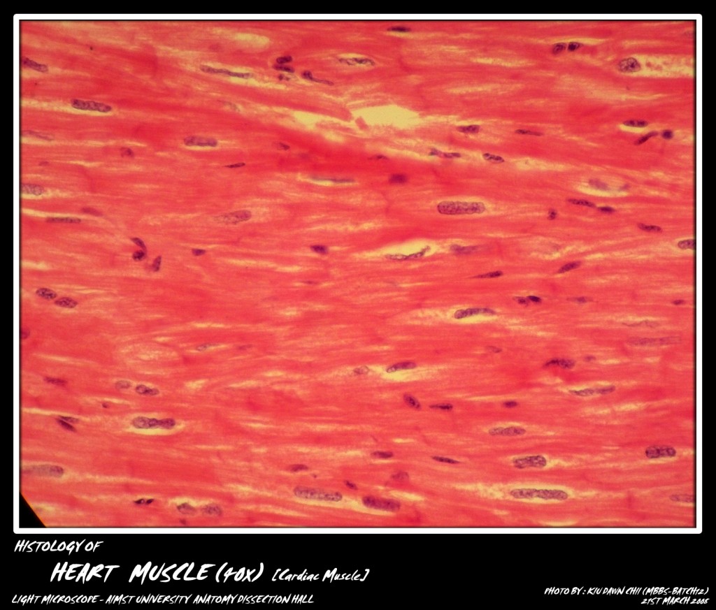
Micro-photograph of Cardiac Muscles under light microscope magnification 40x (intercalated discs can be seen)
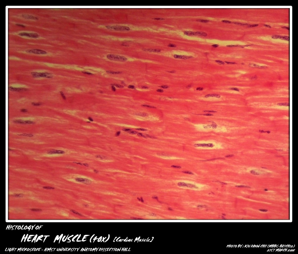
Micro-photograph of Cardiac Muscles under light microscope magnification 40x (intercalated discs can be seen)
Adapted from:http://myaimst.net/mbbsb12/photo/histo/yr1histo/muscle.html
Micro-photograph taken at AIMST University Anatomy Dissection Hall during Histology class, using Canon A40 camera over light microscope.

