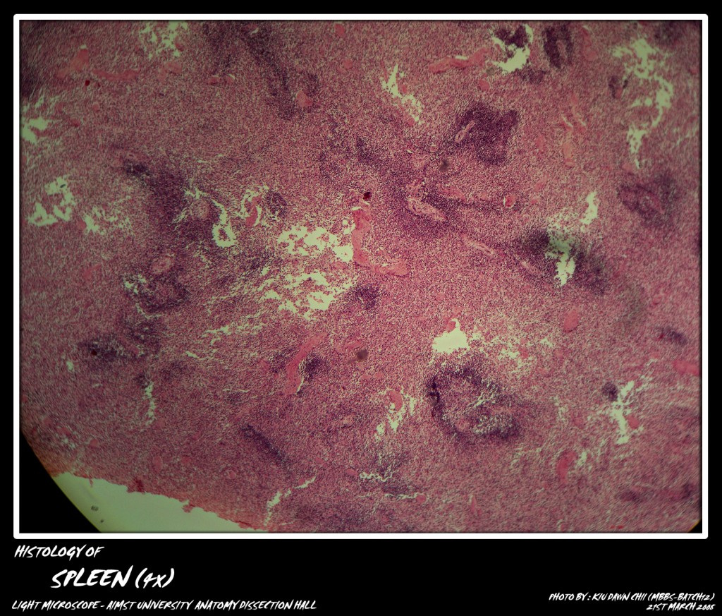
Spleen contains large amount of lymphatic tissue which filters blood instead of lymph. The spleen is surrounded by a connective tissue capsule which in lower animals contains smooth muscle fibers and elastic fibers. The outer surface of capsule is covered by a layer of flattened mesothelium. Trabeculae made up of reticular, elastic and collagenous fibers extend from the capsule and hilus into the splenic pulp. These run in various directions and become continuous with the reticular framework of the organ. There is no differentiation of tissue into cortex and medulla. Instead the spleen is comprised of white pulp and red pulp.
Micro-photograph of spleen (showing white pulp and red pulp) under light microscope magnification 4x
Micro-photograph of spleen (showing white pulp and red pulp) under light microscope magnification 10x
White Pulp: White Pulp consists of lymphatic tissue which forms sheaths around the arterioles. In microscopic sections it is seen in the form of lymphatic nodules usually with an eccentric arteriole. These are known as splenic corpuscles or Maipighian corpuscles. Their peripheral part contains small lymphocytes while the central part/germinal centre has large lymphocytes and plasma cells.
Red Pulp : A loose framework of reticular fibers forms the foundation of red pulp. In the meshes of this framework are many lymphocytes, free macrophages and all the elements of circulating blood i.e. red blood cells, neutrophils and monocytes. It also contains venous sinuses lined by the reticulo endothelial cells. Spleen contains only efferent lymph vessels. “T zone of lymphocytes” is around the perivascular sheath.
Micro-photograph of spleen (showing white pulp and trabecula) under light microscope magnification 10x
Splenic circulation
The tortuous splenic artery divides into 5-7 branches at the hilus of spleen. All these branches enter the spleen through its trabeculae and are named trabecular arteries. Arteriolar branches of trabecular arteries leave the trabeculae and get surrounded by lymphatic nodules. These lymphatic nodules with eccentric arterioles constitute the Malpighian corpuscles or white pulp. The arterioles then enter the red pulp, where they divide into number of straight branches or penicilli. These penicilli are surrounded by reticular cells as well as macrophages and are known as ellipsoids. From the level of ellipsoids the vessel continues as capillaries the fate of which is controversial. According to the ‘closed theory’, the capillaries are continuous with venous sinusoids of the red pulp which convey blood to the trabecular veins and further into tributaries of splenic vein which join to form the splenic vein. This is termed as “closed circulation”. According to the “Open theory”, the capillaries open directly into the red pulp.
The blood from the red pulp returns into the venous circulation through the walls of the “fenestrated venous sinusoids’. The “compromise theory” claims that the splenic circulation is ‘open’ in distended spleen and ‘closed’ in the contracted spleen.
Functions:
1. The reticuloendothelial cells, reticular cells and macrophages are concentrated in the spleen. These cells destroy the worn out red blood cells, white blood cells and micro-organisms.
2. The activated lymphocytes and antibodies produced in the spleen help in various immune/defense mechanisms of the body.
Adapted from : http://myaimst.net/mbbsb12/photo/histo/yr1histo/lymphoidorgan.html
Micro-photograph taken at AIMST University Anatomy Dissection Hall during Histology class, using Canon A40 camera over light microscope.

