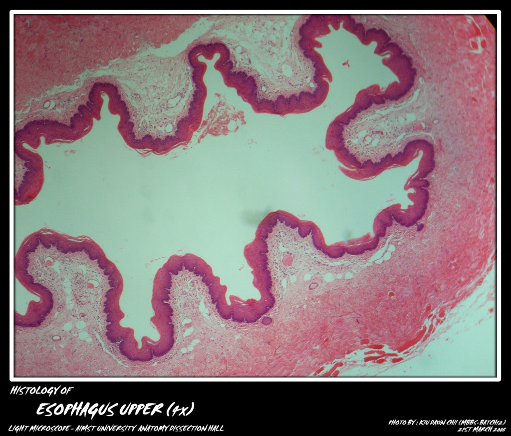
The esophagus is a muscular tube that rapidly propels the food from pharynx into the stomach. It is about 25 cms long. Its wall includes all the layers included in the general plan of G.I.T.
The mucous membrane is thrown into longitudinal folds when empty. These folds become smooth as the bolus of food passes through the esophagus. The epithelium is stratified squamous non. keratinised in character and protective in function. The lamina propria sends papillae into the epithelium. The muscularis mucosae is indistinct at the beginning of esophagus, but becomes distinct lower down. It is made up of longitudinal layer of smooth muscle fibers.
The submucosa contains oesophageal glands. These are mucus secreting glands with acini which are round or oval in shape. The lining cells are truncated columnar with flattened peripheral nuclei. The cytoplasm is lightly stained and contains mucigen droplets.
The muscularis externa has striated muscle fibers in upper third, mixed (striated and smooth) muscle fibers in the middle third and smooth muscle fibers in the lower third of esophagus. The outermost layer is the adventitia which is made up of loose connective tissue with capillaries and nerves.
Micro-photograph of Stratified Squamous Epithelium of Esophagus Lower under light microscope magnification 40x
Adapted from: http://myaimst.net/mbbsb12/photo/histo/yr1histo/esophagus.html
Micro-photograph taken at AIMST University Anatomy Dissection Hall during Histology class, using Canon A40 camera over light microscope.

